- Shoulder
- Anatomy
- Pathology
- Interventions
- Block of suprascapular nerve
- Infiltration bicipital recess - long head of biceps tendon
- Infiltration subacromial subdeltoid bursa
- Injection AC-Joint
- Injection AC-Joint after resection
- Injection glenohumeral joint lateral and medial approach
- Injection trapezius or levator muscle
- Needling calcific tendinitis supraspinatus tendon
- Scapulothoracic bursa Injection
- Elbow
- Hand
- Anatomy
- Pathology
- Fingers
- dorsal
- Crystal arthropathy
- Distal phalanx fracture
- Epithelial cyst / ganglion cyst of the finger
- Foreign body (rose thorn, glass)
- Giant cell tumor finger
- Gouty arthropathy
- Mallet Finger (Baseball finger)
- Mucoid / ganglion cyst DIJ
- Partial tear extensor tendon / mechanism 5th finger
- Tear Abductor digiti minimi
- Ulnar collateral ligament tear / Stener lesion
- Venous aneurysm malformation PIP
- [PsA] Erosions
- [PsA] Inflammation finger, enthesitis, tendinitis extensor dig. tendon
- [PsA] enthesitis extensor dig. tendon
- [RA] Cartilage measurement in rheumatoid arthritis
- [RA] Erosions and synovitis
- [RA] Tenosynovitis of the extensor / flexor tendons
- [SpA] Finger DIJ Synovitis
- finger joint prosthesis
- palmar
- A2 pulley tear
- Complete tear superficial digital flexor tendons, partial tear deep flexor tendon
- Degenerative osteoarthritis
- Foreign body (glass splinter) finger
- Prosthesis TMC-Joint
- Tendovaginitis stenosans flexor tendons
- Tenosynovitis of the flexor tendons from overuse
- [CPPD] MCP joints arthropathy
- [PsA] Enthesitis of the flexor tendon DIJ
- [PsA] Inflammation finger
- [RA] Effusions, grade 1–3
- [RA] Synovitis MCP joints
- [RA] Tears of the flexor tendons, tenosynovitis
- [RA] Tenosynovitis of the extensor / flexor tendons
- [SpA] Finger DIJ Synovitis
- [SpA] Flexor tendons tenosynovitis
- dorsal
- Palm
- Wrist
- dorsal
- Carpal boss
- De Quervain tendinopathy
- Ganglion cysts
- Osteomyelitis and soft tissue infection after MC-fracture and dorsal plate
- Posttraumatic instability DRUG
- Radiodorsal ganglion cyst of the wrist (SL-ligament)
- Tear radio- and lunotriquetral ligament, ganglion cyst wrist
- [CPPD] Calcifications extensor carpi ulnaris tendon
- [CPPD] Ganglion cyst extensor carpi ulnaris tendon
- [CPPD] Osteophytes radiocarpal joint
- [CPPD] active erosion
- [RA] Synovitis of the wrist
- [RA] active erosion capitate
- [RA] rheumatic nodule
- accessory muscle extensor tendons
- palmar
- dorsal
- Fingers
- Interventions
- Aspiration and puncture of ganglion cyst
- Aspiration of peritendinous effusion
- Carpal tunnel syndrome perineural infiltration median nerve
- Infiltration MCP joint in Psoriatic arthritis
- Infiltration PIJ and DIJ
- Infiltration flexor tendons tenosynovitis in plane / out of plane
- Injection STT Joint
- Injection TMC Joint
- Injection ganglion cyst / mucoid cyst DIJ
- Injection of the forth extensor tendon compartment
- Injection trigger finger
- Injection wrist joint doppler
- Puncture and injection FPL tendon pulley ganglion cyst
- Hip
- Anatomy
- Pathology
- Interventions
- ACP InjectionTuber ischiadicum / hamstrings
- Arthrocentesis hip
- Infiltration Meralgia paraesthetica
- Injection acetabular labrum with ACP
- Injection gluteal tendons hyaluronic acid / ACP
- Injection hip
- Injection paralabral ganglion cyst
- Injection trochanteric bursa
- Puncture and infiltration of iliopsoas bursitis
- Puncture trochanteric bursa / hematoma after tear
- Trigger point injection gluteal region doppler
- Knee
- Foot
- Anatomy
- Pathology
- Ankle
- anterior
- lateral
- medial
- plantar
- posterior
- Achilles tendon
- Achilles tendinopathy and bursitis from overuse
- Achilles tendinopathy with calcification
- Complete tear Achilles tendon
- Fluoroquinolone-induced Tendinopathy of the Achilles tendon
- Non-insertional achilles tendinopathy
- Partial tear and tendinopathy of the Achilles tendon
- Retrocalcaneal bursitis
- Subcutaneous calcaneal bursitis
- Tear medial head of gastrocnemius
- Tear of the plantaris tendon
- [PsA] Enthesiopathy Achilles tendon
- [SpA] Achilles tendon enthesitis
- Ganglion cyst ankle joint
- Tear medial head gastrocnemius, hematoma
- Achilles tendon
- Midfoot
- Toes
- dorsal
- Activated osteoarthritis MTP Joint
- Intraarticular calcification MTP Joint
- Morton neuroma
- Proximal phalanx fracture toe
- [CPPD] Calcifications MTP Joint
- [Gout] Synovitis / Tophus MTP Joint
- [PsA] Metatarsophalangeal joint synovitis and periarticular inflammation
- [RA] MTP joint synovitis
- [RA] Metatarsophalangeal joint effusion
- plantar
- dorsal
- Ankle
- Interventions
- Spine
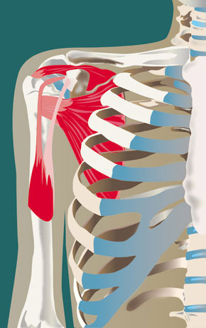
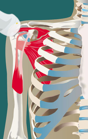
Anatomy of the Shoulder
Pathology of the Shoulder
Interventions in the Shoulder Region
- Block of suprascapular nerve
- Infiltration bicipital recess - long head of biceps tendon
- Infiltration subacromial subdeltoid bursa
- Injection AC-Joint
- Injection AC-Joint after resection
- Injection glenohumeral joint lateral and medial approach
- Injection trapezius or levator muscle
- Needling calcific tendinitis supraspinatus tendon
- Scapulothoracic bursa Injection
Color Legend
Red: Inflammatory
Blue: Degenerative/Trauma/Tumor
Green: Crystals
Tears: Supraspinatus
Patient Positioning
Patient seated on a revolving stool. Position one: The palmar side of the hand on the superior aspect of the iliac wing, elbow posterior. Position two: The dorsum of the hand over the opposite back pocket, which leads to an internal rotation. The supraspinatus is directed more anteriorly.
Probe Positioning
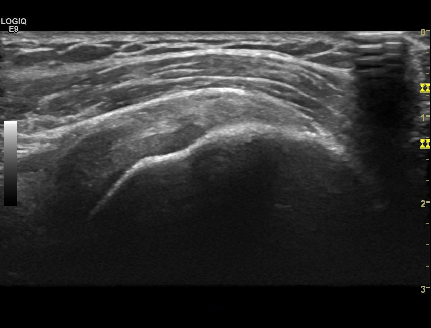
The intraarticular portion of the long head of biceps is found easily medial - rotate the probe to get its long axis. The long head of biceps and the supraspinatus tendon run in parallel. Then shift the transducer a little cranially and posteriorly over the supraspinatus tendon - this is the long axis view of the tendon. Scan the entire visible portion of the tendon.
Partial-thickness articular-sided tear of the supraspinatus tendon
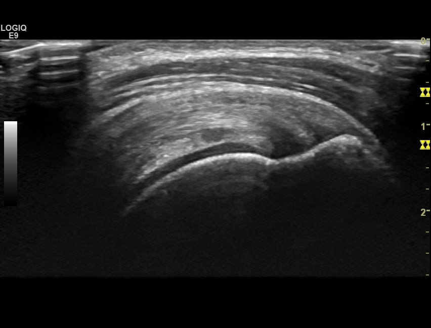
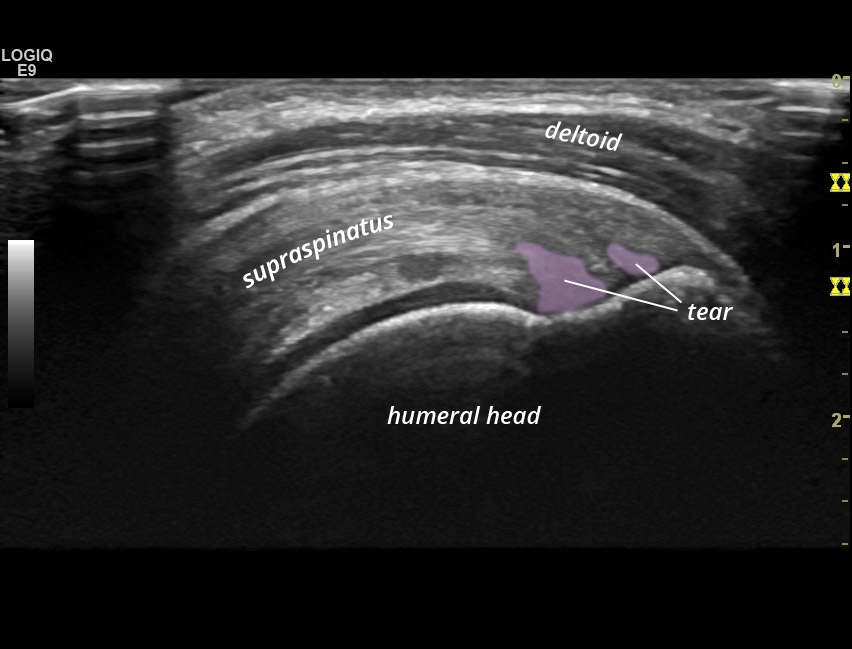
Partial-thickness articular-sided tear of the supraspinatus tendon
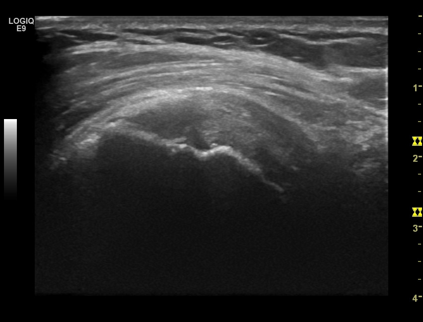
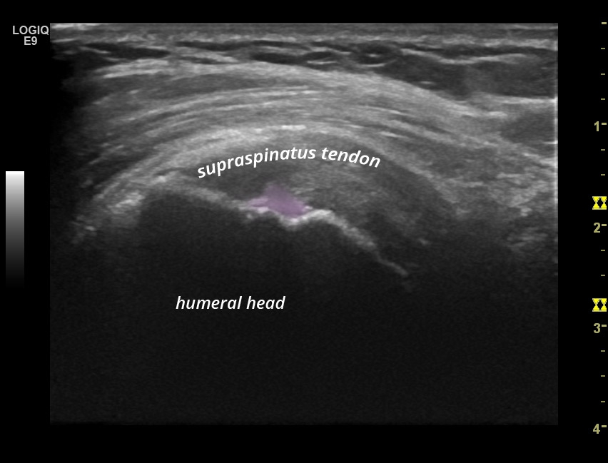
Articular sided partial thickness supraspinatus tendon tear
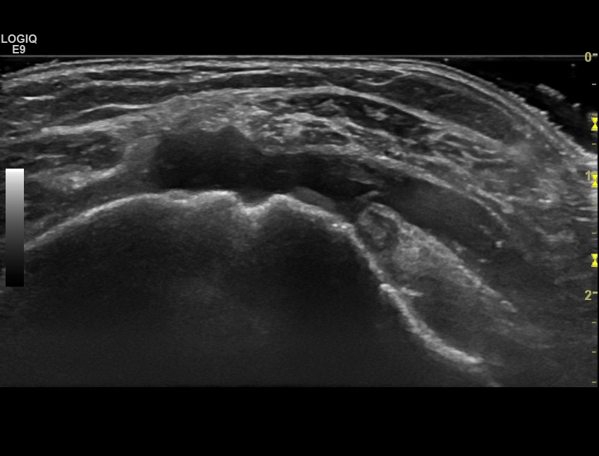
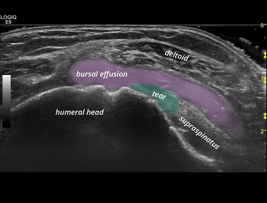
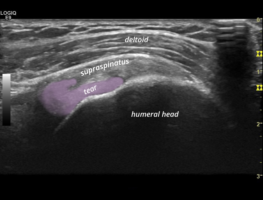
Full thickness tear of the supraspinatus tendon, subacromial subdeltoid bursitis
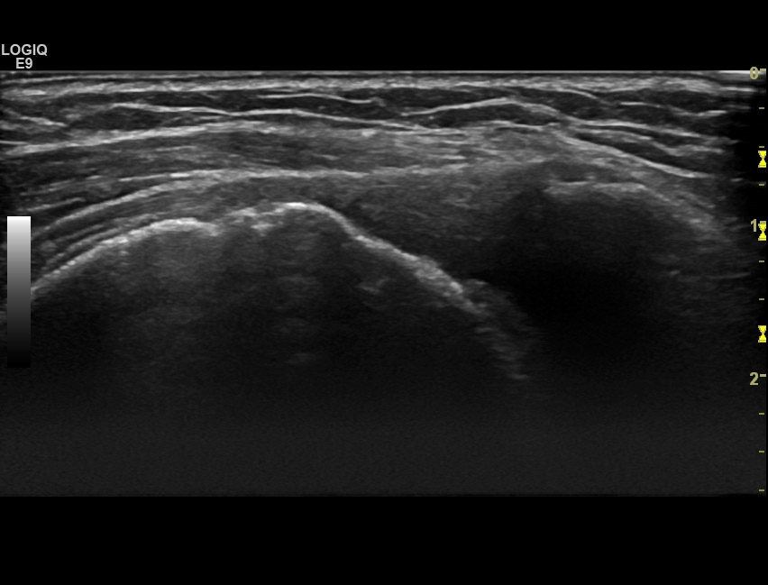
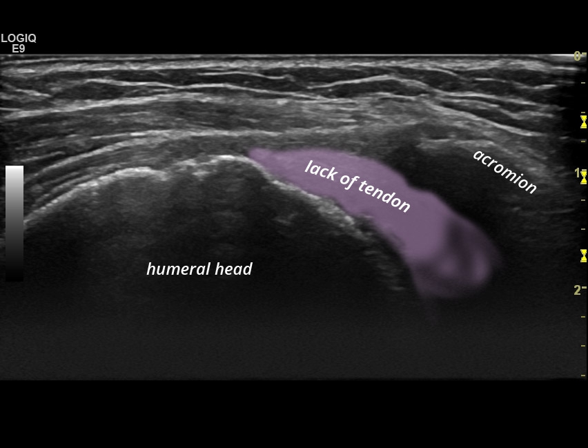
Complete tear of the supraspinatus tendon with non-visualization of the tendon and decreased acromiohumeral distance
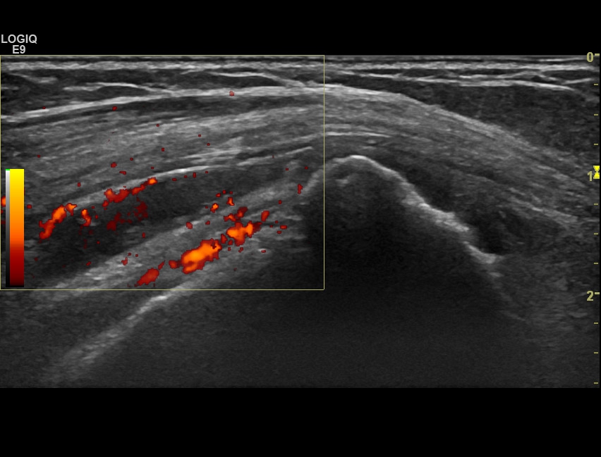
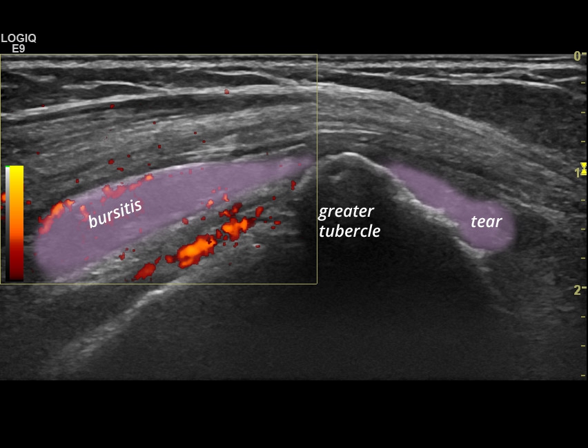
Subacromial subdeltoid bursitis with bursal effusion and positive dopplersignal, total thickness tear of the supraspinatus tendon.

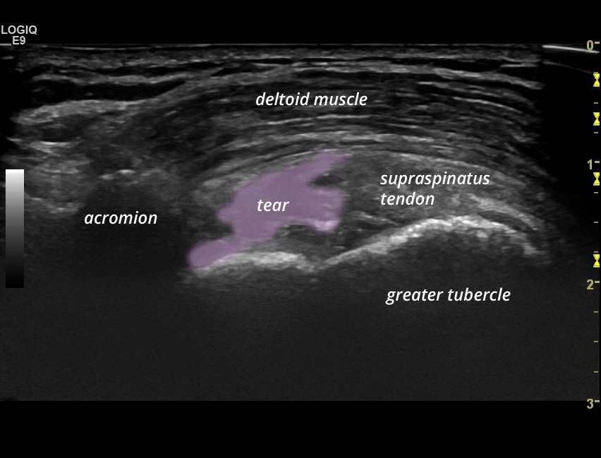
Full thickness tear of the supraspinatus tendon with reduced acromiohumeral distance
![]()
© 2025 Rheuma Schweiz
Privacy Preference
Here you will find an overview of all cookies used. You can give your consent to whole categories or display further information and select certain cookies.
Accept all Save Accept only essential cookies BackEssential
Essential cookies enable basic functions and are necessary for the proper function of the website.
Show Cookie Information| Name | cookieconsent_status |
|---|---|
| Provider | Owner of this website |
| Purpose | Saves the settings of the visitors selected in the cookie box. |
| Cookie Name | cookieconsent_status, cookieconsent_statistik, cookieconsent_medien |
| Cookie Expiry | 1 year |
| Name | Google Tag Manager |
|---|---|
| Provider | Google LLC |
| Purpose | Google cookie to control advanced script and event handling. |
| Privacy Policy | https://policies.google.com/privacy?hl=en |
| Cookie Name | _ga,_gat,_gid |
| Cookie Expiry | 2 years |
Statistics
Statistics cookies collect information anonymously. This information helps us to understand how our visitors use our website.
Show Cookie Information| Accept | |
|---|---|
| Name | Google Analytics |
| Provider | Google LLC |
| Purpose | Cookie by Google used for website analytics. Generates statistical data on how the visitor uses the website. |
| Privacy Policy | https://policies.google.com/privacy?hl=en |
| Cookie Name | _ga,_gat,_gid |
| Cookie Expiry | 2 years |
External Media
Content from video platforms and social media platforms is blocked by default. If External Media cookies are accepted, access to those contents no longer requires manual consent.
Show Cookie Information| Accept | |
|---|---|
| Name | Vimeo |
| Provider | Vimeo |
| Purpose | Used to unlock Vimeo content. |
| Privacy Policy | https://vimeo.com/privacy |
| Host(s) | player.vimeo.com |
| Cookie Name | vuid |
| Cookie Expiry | 2 years |
Supported by